
Categorised under:
Interventional Radiology
>
Imaging equipment
>
Ultrasound
Imaging
>
US Scanners
>
Compact
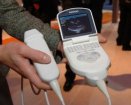
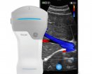
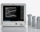
From Zonare
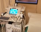
According to the manufacturer, the Zone Sonography, developed by ZONARE Medical Systems, is a new approach to data acquisition and image formation. Zone Sonography acquires larger quantities of data very quickly in relatively few “zones” and processes the data at the speed of light—in other words, the speed of the processor. The throughput is therefore substantially faster and there is more time for a variety of advanced processing options while still delivering extremely high image quality.
Since it is implemented almost entirely in software, Zone Sonography systems are smaller, lighter and less expensive than traditional systems offering equivalent image quality—yet deliver performance and flexibility that far exceeds any other ultrasound technology.
Your opinion matters to others - rate this device or add a comment


Did you know you can Register for FREE with this website?
Registration gives you full access to all of the features of WhichMedicalDevice. Find out more ...
WhichMedicalDevice is a FREE resource created by clinicians for clinicians.
Registration is free and gives you unlimited access to all of the content and features of this website.
Find out more...Registration is free and gives you unlimited access to all of the content and features of Which Medical Device. Find out more...
Which Medical Device is a community of clinicians sharing knowledge and experience of the devices and procedures we use on a daily basis. We ask that our members register with us so that we can maintain the unbiased and independent nature of our content. Registration is quick and free.
We do not make your details available to any third parties nor do we send unsolicited emails to our members. You can read our Privacy Policy here.