
Categorised under:
Interventional Radiology
>
Gastrointestinal
>
T Fasteners, gastropexy
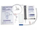
In summer 2007 Boston Scientific discontinued the Brown-Müller T-fastener, which has been a land-mark device since its first description in 1986(1). A French clone of this manufactured by Balt and distributed in the UK by Pyramed, Basingstoke, UK became available in October 2007. The main difference between the two sets is that the Harpon is supplied with 3 T-fasteners as opposed to 4. Originally the Harpon had two metal bars to be crimped onto the suture, but is now supplied with a single larger crimp.
In summer 2007 Boston Scientific discontinued the Brown-Müller T-fastener, which has been a land-mark device since its first description in 1986 (1). A French clone of this manufactured by Balt and distributed in the UK by Pyramed, Basingstoke, UK became available in October 2007. The main difference between the two sets is that the Harpon is supplied with 3 T-fasteners as opposed to 4. Originally the Harpon had two metal bars to be crimped onto the suture, but is now supplied with a single larger crimp.
Insertion of feeding tubes directly through the skin is almost exclusively performed by radiologists and has been termed radiologically inserted gastrostomy, RIG or percutaneous radiological gastrostomy PRG (2). It allows primary placement of low-profile devices and transgastric jejunostomy tubes (3).
The philosophy behind gastropexy is that while pushing the tube through the abdominal wall it may displace the stomach and end up in the peritoneal cavity. Gastropexy reduces the risk of peritoneal displacement by fixing the stomach to the anterior abdominal wall (gastro - stomach, pexy - fixation).
Most tubes used for RIG have a balloon as an internal fixator, alternatives are tubes using a Cope-loop type locked PIG-tail (Tilma, Cook, Letchworth, UK) or a foldable mechanical bumper (e.g., Entristar, Tyco, Gosport, UK), but the former have a relatively high rate of occlusion and displacement and the latter require large insertion tracks (4, 5).
The disadvantage of balloons is their risk of rupture and their loss of volume over time by osmosis of water through the silicone membrane. For this reason tube manufacturers recommend changing the water in the balloon on a weekly basis and changing the tube every 3 months.
How long gastropexy should be left in situ remains under debate. From infection control and pain point of view it would be desirable to remove the sutures as soon as possible; however, in our group of malnourished cancer patients undergoing chemotherapy we have seen 2 cases where the stomach did not adhere to the anterior abdominal wall, even after 2 weeks, resulting in catheter displacement after cutting of the gastropexy sutures.
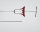
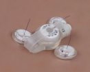
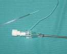
The T-fasteners consist of a small metal bar attached to a nylon suture (Fig. 1). The bar is inserted into the tip of a slotted needle (Fig. 2), which is inserted under local anaesthesia and fluoroscopic control through the skin into the inflated stomach. After confirmation of correct position by injection of contrast, the T-bar is ejected from the needle tip into the gastric lumen with the use of a steel pusher (Fig. 3). The needle is removed leaving the T-fastener behind, and gentle traction apposes the stomach to the anterior abdominal wall. On the outside a cotton wool bud is mounted on the suture below a disc and a small metal tube. These are slid down to skin level at which point the metal tube is crimped to the suture creating the external fixation. The prongs of even large forceps are not strong enough to crimp the metal tube and this is better done by compressing the tube with the handles behind the hinge (Fig. 4). The needle is used to puncture between the sutures and place a stiff guide wire. (Fig. 5). After dilatation an appropriate sheath is inserted, which causes significant traction on the sutures (Fig. 6). The G-tube is then placed through the sheath and the outer fixator adjusted to sit snug but not tight against the skin (Fig. 7).
For removal of the sutures these are simply cut at skin level below the cotton wool bud under gentle traction. In theory the internal metal bar and the inner suture displace into the stomach, in practice they are often seen still in situ after several weeks.
It is unclear how many sutures are really required. For insertion of balloon catheters a rule of thumb is creating a track 4Fr. larger than the actual tube size, e.g., a 16Fr. track/peel-away sheath for a 12Fr. balloon catheter. Obviously, larger tubes exert greater force on the stomach and the T-fasteners, but 3 sutures are perfectly adequate for tubes up to 14Fr. and the required 18Fr. peel-away sheath. Small bore loop-retained catheters are safely inserted using only 1 or 2 T-fasteners, but in our practice these tubes have performed inferior to balloon catheters. The Entristar, although nominally a 12Fr. tube, requires a 22Fr. track due to the large folding retainer and careful serial dilatation is required for these.
SEE THE VIDEO OF GASTROSTOMY INSERTION FOR THE USE OF THIS TYPE OF GASTROPEXY.
The main alternatives are the Cope-sutures from Cook. The packs contain 2 T-fasteners fashioned from flexible guide wires and are in theory less traumatic to the stomach. The suture ends in a needle and the instructions for use suggest suturing the T-fastener to the skin. In practice this causes confusion of the nurse trying to remove these and sometimes they are left in situ altogether resulting in bad stoma infection.
A number of more constructive approaches have been tried, e.g., attaching the sutures to the outer fixation disc of the catheter, but this prevents rotation of the tube. The most practical solution was suggested to me by Dr. D. Fay from Bath, who applies an adhesive stoma disc around the tube to which the sutures are applied.
Hans-Ulrich Laasch
Christie Hospital, Manchester, UK

Did you know you can Register for FREE with this website?
Registration gives you full access to all of the features of WhichMedicalDevice. Find out more ...
WhichMedicalDevice is a FREE resource created by clinicians for clinicians.
Registration is free and gives you unlimited access to all of the content and features of this website.
Find out more...Registration is free and gives you unlimited access to all of the content and features of Which Medical Device. Find out more...
Which Medical Device is a community of clinicians sharing knowledge and experience of the devices and procedures we use on a daily basis. We ask that our members register with us so that we can maintain the unbiased and independent nature of our content. Registration is quick and free.
We do not make your details available to any third parties nor do we send unsolicited emails to our members. You can read our Privacy Policy here.