


Categorised under:
Orthopaedics
>
Oncology
>
Extendible prostheses
Orthopaedics
>
Paediatric
>
Paediatric oncology
Reconstruction of massive bone defects after the resection of primary bone tumours in children remains a challenge for orthopaedic oncology surgeons. Custom made implants represent one solution to this problem, but need to accommodate growth. As a result, several designs of growing implants have been developed over the years comprising many different and ingenious lengthening mechanisms. Since the 1980s designs from Stanmore Implants Worldwide have been lengthened by the insertion of ball bearings, metal C-shaped spacers of varying length, and by a worm-wheel screw mechanism lengthened by inserting an Allen key. All of these systems had the disadvantage that the patient required an operation for the implant to be lengthened, with the attendant risks of infection in particular.
Reconstruction of massive bone defects after the resection of primary bone tumours in children remains a challenge for orthopaedic oncology surgeons. Custom made implants represent one solution to this problem, but need to accommodate growth. As a result, several designs of growing implants have been developed over the years comprising many different and ingenious lengthening mechanisms. Since the 1980s designs from Stanmore Implants Worldwide have been lengthened by the insertion of ball bearings, metal C-shaped spacers of varying length, and by a worm-wheel screw mechanism lengthened by inserting an Allen key. All of these systems had the disadvantage that the patient required an operation for the implant to be lengthened, with the attendant risks of infection in particular.
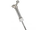
The non-invasive prosthesis represents a significant advance in this technology. The core is a motor which spins at high revolutions (around 13,000 rpm) when placed inside a radiofrequency coil and acts through a gearing mechanism to apply a lengthening force to the limb. This technology is the result of many years of research by the Stanmore team.
Considerations include the length of bone to be resected and the predicted growth of the child. The length of the internal mechanism means that the system may not be suitable if the resection is very short. Furthermore, if the patient is approaching skeletal maturity, an acute lengthening (of 1 to 2 cm) may suffice. The lengthening required by the device depends on which physis is lost. Typically, in the distal femur, there will be loss of the distal femoral physis (0.9cm per year), and the instrumentation of the proximal tibial physis reduces growth in it to around 70% of normal, even with a smooth stem placed through the physis. As a result, in very young children, the knee on the operated side may ultimately lie slightly lower than the other.
Once the tumour has been resected, the implant can be inserted. When replacing the distal femur the author’s preference is to cement the femoral stem of the implant in the belief that this leads to easier revision than the attempted removal of an ingrown cementless stem. Usually, this implant would have a hydroxyapatite collar to encourage bone ongrowth onto the implant and discourage loosening. On the tibial side, the articular surface can be removed using a zero degree cutting jig, and the tibial component inserted. This can be cemented onto the surface of the tibia if care is taken to keep cement out of the physis.
Particular technical points to note are:
It is recommended that after implantation, the device is activated briefly to ensure proper function. Lengthening involves placing the limb inside a radiofrequency coil which spins the motor and lengthens the implant at the rate of 1mm in 4 minutes. The machine has two settings – A and B. Usually, a distal femoral implant will require the A setting for lengthening and B to reverse the process. When activated, the motor can be heard spinning inside the implant and makes a noise said to be like running water. This can be heard with a stethoscope or by listening carefully to the limb. Selecting the alternative setting reverses the motor and allows the limb to be shortened. Alternative use to lengthen limbs in skeletally mature patients with shortening after revision of failed reconstructions has been described (Sewell et al).
I have had excellent results with this implant, and although failure of the lengthening mechanism has been described these are unusual and the motor can sometimes be kick-started using a higher energy coil. I have had no failures in this regard. To see patients having lengthenings in the clinic which are painless and quick is little short of miraculous.
Although some surgeons prefer different approaches to this clinical problem (such as rotationplasty) competing devices include the Repiphysis (Wright Medical, previously known as Phenix, Phenix-medical, Paris, France). This device has a compression spring set in polyethylene within the implant. To lengthen the device a radiofrequency coil heats up a receiver coil which warms up the polyethylene allowing the spring to act. There is at least one report of failure of the lengthening mechanism (1). Another option is the MUTARS BioXpand device (Implantcast, Buxtehude) which in theory uses callus distraction to lengthen to limb, increasing the length of the host bone segment, rather than the length of the implant.
This is an excellent device which has transformed the lives of children undergoing endoprosthetic reconstruction.
To see someone undergoing limb lengthening with this device, look at Alex Dawson’s story on Jimmyteens.tv
For a review of growing implant technology try this open source article by Nystrom and Morcuende.
There are no currently similar devices - Click here to suggest a device

Did you know you can Register for FREE with this website?
Registration gives you full access to all of the features of WhichMedicalDevice. Find out more ...
WhichMedicalDevice is a FREE resource created by clinicians for clinicians.
Registration is free and gives you unlimited access to all of the content and features of this website.
Find out more...Registration is free and gives you unlimited access to all of the content and features of Which Medical Device. Find out more...
Which Medical Device is a community of clinicians sharing knowledge and experience of the devices and procedures we use on a daily basis. We ask that our members register with us so that we can maintain the unbiased and independent nature of our content. Registration is quick and free.
We do not make your details available to any third parties nor do we send unsolicited emails to our members. You can read our Privacy Policy here.