
Categorised under:
Interventional Radiology
>
Imaging equipment
>
Ultrasound
Imaging
>
US Scanners
>
Compact
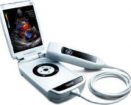
The Zonare ultrasound system is a fully-fledged ultrasound machine packed into a compact portable 'scan engine'. The machine differs from conventional portable machines and larger cart based systems in many ways. First of all do not be fooled by its diminutive size. The technology involved (more later) means that this machine is fully capable of high resolution B mode scanning with the addition of compound imaging and tissue harmonics. It is also capable of spectral Doppler, colour Doppler and power Doppler.
The Zonare ultrasound system is a fully-fledged ultrasound machine packed into a compact portable ‘scan engine’. The machine differs from conventional portable machines and larger cart based systems in many ways.
First of all do not be fooled by its diminutive size. The technology involved (more later) means that this machine is fully capable of high resolution B mode scanning with the addition of compound imaging and tissue harmonics. It is also capable of spectral Doppler, colour Doppler and power Doppler.
We managed to borrow a machine for two weeks and put it through its paces. We were looking for a machine with all the features of our standard departmental ultrasound machines (Philips IU22), but that could be easily used in the interventional room and operating theatres for interventional procedures such as nephrostomy, PCNL, RF ablation, PTC and intraoperative tumour localisation. We also needed a machine with sensitive colour Doppler for post organ transplant vascular assessments. Our current problem is that the machine we use in intervention is large, poorly manoeuvrable and of limited ability. Needles are difficult to see, tissue contrast is poor and RF generators interfere with the image. We also need a machine that can easily be moved up one floor to the operating theatres at short notice without going in the lift!
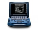
The Zonare Z.one is essentially the portable ‘scan engine’. This can be mounted on the cart to give a larger screen, keyboard, easier access to controls, somewhere to put the probes and jelly. The scan engine can be operated to full capability when detached from the cart and easily carried in one hand.
This is a little difficult to explain and to be honest I only understand the basics of how it works. The Zonare machine uses a fundamentally different mechanism of data acquisition and image formation to other ultrasound machines. Data is acquired in ‘zones’. Each new zone that is acquired has data that overlaps with the previous zone. This data is held in Channel domain memory. Areas of the image with a low signal to noise ratio have a larger amount of overlap with data from other zones leading to uniformity of signal detection. Zone sonography treats all of the received echoes as useful information. This is done by analysing returning echoes from each pixel and adjusting them to compensate for the variation in intensity of the transmit wave. This creates an effective focus at every pixel in the image. This explains why there is no focus control on the Zonare machine as all pixels are ‘focussed’.
The other advantage of zone sonography is the short sequence of acquisition events to produce an image frame. This allows plenty of ‘free time’ in the acquisition cycle to acquire other modes without reducing the B mode quality.
Zonare claim that their technology moves ultrasound from a speed of sound constrained environment to a computational speed environment. Thus with Moore’s Law stating that computational power will roughly double every eighteen months (and it still is) there will be continual improvements in this machine’s speed and capability.
Our machine had clearly been around quite a few hospitals but seemed to be standing up well. The only minor problem we had was it required a reasonable amount of force to re engage the scan engine with the cart after portable use.
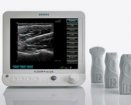
The cart itself is sturdy but light and easily manoeuvred. There are plenty of slots for the many available probes. The screen is a large LCD that can be swivelled in any direction. The scan engine is easily released by a catch just anterior to it. A useful feature is that it can be released and attached whilst turned on. Probes are attached on the right of the scan engine and again can be ‘hot swapped’ which does save time.
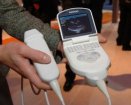
The scan engine is sturdy but easy to carry in one hand. Its best placed on a trolley or stool for portable scanning as it is quite heavy if using for a prolonged time.
Probes
C5-2 curved array, abdominal, obstetrics, intervention. (2 harmonic frequencies)
E9-4 endovaginal probe, obstetrics, (1 harmonic frequency)
L10-5 linear array, small parts, peripheral vascular, (1 harmonic frequency)
L8-3 linear array, peripheral vascular, small parts, transverse needle guide built in, (2 harmonic frequencies)
P10-4 phased array, neonates, paediatrics, (1 harmonic frequency)
Power up is quick. (I get very irritated by machines that seem to take forever to boot when you are desperate to get on with a complex case.) Patient demographics are entered in a standard way. Select the probe you need by simply plugging it in. Menu options are accessed by the ‘rocker pad’ on the left (see images). The keyboard is located below the display, which I initially found irritating (probably because the machine I am used to is the opposite). The keyboard is actually rarely used once you are scanning. There are also several ‘soft keys’ beneath the display whose function changes depending upon the selected mode.
The controls are fairly standard and easy to find your way around. It’s initially a little disconcerting to have no focal zone adjustment. There is a very useful ‘optimize’ button that ‘does what it says’ to the image, adjusting several parameters without having to enter any menus.
The monitor is a flat panel LCD (1280x1024) mounted on a movable arm. The actual image takes up less than 1/3 of the display but I understand this will be user configurable in the near future. The remainder of the display contains patient demographics, drive capacity, recent measurements and your last three stored images.
Images are stored onto hard disc but can also be stored on USB flash memory if needed. PACS linkage is standard. Images can also be exported onto USB flash memory for importing into teaching collections and PowerPoint. One thing to note is that they do not get a file extension. The still images are .TIF format and the videos are QuickTime .MOV files. These file extensions need to be added manually once copied onto your computer.
The scan engine is easily removed from the cart. All the same functions can be accessed via the menu system. The screen is fairly small but is of very good resolution and we did not find that this compromised scanning.
We have taken a variety of images of some of our cases over the two weeks we had the machine (see images) Some are shown with comparable images of the same patient scanned with a Philips IU22 machine which is our current ‘gold standard’.
Grey scale imaging was superb with excellent tissue differentiation. Compound imaging appeared to give a slightly over smoothed image with some probes and I mostly used it turned off. Others who used the machine disagreed with me on this and preferred the compound imaging. Tissue harmonics were good and I found them especially helpful with larger patients and renal work.
The machine showed very good Doppler sensitivity. One of my colleagues managed to demonstrate a type 3 endoleak with this machine that he had previously only been able to see with the IU 22.
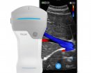
The 10/4 sector probe has a very small footprint useful for paediatrics and some interventional work (see images). The 10/4 linear was excellent for vascular and ‘small parts’ such as thyroid and testes. We found the 5/2 MHz curvilinear probe best for general work and abdominal intervention.
I performed 2 renal RF ablations and a colleague performed a liver RF ablation using the Zonare for guidance. In all cases the needle was very easy to see compared with our standard interventional machine and there was no interference from the RF generator.
Unfortunately we did not manage to try out all the probes including the hockey stick and endocavity probes
When I first saw this machine I was a little sceptical that it was a fully capable scanner in such a small package. 2 weeks of use both within the ultrasound department, in theatres, in the IR room and on the wards have proven that it is. The comparison pictures we have taken also add credence to this. I am not aware of another machine on the market that can be both cart based and portable yet offer these features.
Dr Philip J Haslam
Consultant Interventional and Uroradiologist
No conflict declared.


Did you know you can Register for FREE with this website?
Registration gives you full access to all of the features of WhichMedicalDevice. Find out more ...
WhichMedicalDevice is a FREE resource created by clinicians for clinicians.
Registration is free and gives you unlimited access to all of the content and features of this website.
Find out more...Registration is free and gives you unlimited access to all of the content and features of Which Medical Device. Find out more...
Which Medical Device is a community of clinicians sharing knowledge and experience of the devices and procedures we use on a daily basis. We ask that our members register with us so that we can maintain the unbiased and independent nature of our content. Registration is quick and free.
We do not make your details available to any third parties nor do we send unsolicited emails to our members. You can read our Privacy Policy here.